Articular cartilage is a highly specialized connective tissue that is covering the ends of long bones. These tissues enable the smooth movement of joints. Articular cartilage consists mainly of a strongly hydrated matrix of collagen type II, and of proteoglycans such as chondroitin sulfate. Osteoarthritis is a common disease progressing appearing as a result of false posture, increased body weight, injuries, and increased average age of the population leading to pronounced degradation of articular cartilage. Next to the apparent cartilage damage, osteoarthritis is frequently accompanied by severe pain, and corresponding movement limitations.
Consequently, non- or minimally-invasive diagnostic tools enabling early recognition of the disease onset are demanded facilitating timely treatment of any cartilage alteration for promoting recovery rates, as has successfully been shown in a variety of animal models where such tools have readily proven their utility during successive treatments, as well as for post treatment evaluation.
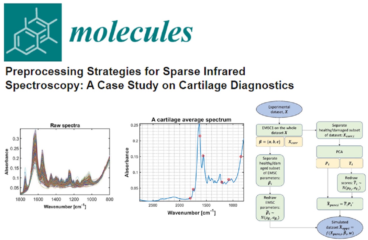
For the evaluation of localized alterations of the cartilage matrix, the likewise localized evaluation of the cartilage matrix integrity is required, which is routinely executed via visual observation and manual probing of the cartilage elasticity during arthroscopy. However, if additional information in molecular detail could be obtained – such as via in vivo arthroscopic mid-infrared (MIR) spectroscopy – an additional dimension of information is provided enabling a more detailed cartilage damage evaluation.
Read the full article on Biophotonics.World‘s website to find out how the research of ULM University will impact on the diagnosis methods for joint cartilage damage – Diagnosing Cartilage Degeneration via Mid-Infrared Spectroscopy: The future of Molecular Arthroscopy?