Detecting Early Cartilage Damage
Which cases MIRACLE device can be used?
MIRACLE can be used for clinical cases where patients are recommended to undergo arthroscopy and the surgeon needs to assess the quality of cartilage lesion to determine whether further action has to be taken.
Arthroscopy is a minimally invasive surgery procedure on a joint in which an evaluation and eventually treatment of cartilage damage is performed. Patients can be referred to arthroscopy due to trauma accident, diagnosis based on X-ray, MRI and symptoms or inconclusive diagnosis. For example, MRI is usually performed before surgery but it may overestimate or underestimate the severity of cartilage lesion, which can be clarified during arthroscopy. The arthroscopy procedure is not performed as health check-up or based only on symptoms.
MIRACLE device has been developed to focus on knee arthroscopy. However, it is possible to miniaturise the developed probe, further enabling the use of the device on smaller joints. The most promising application for MIRACLE is post-traumatic assessment due to sport or accident injury.

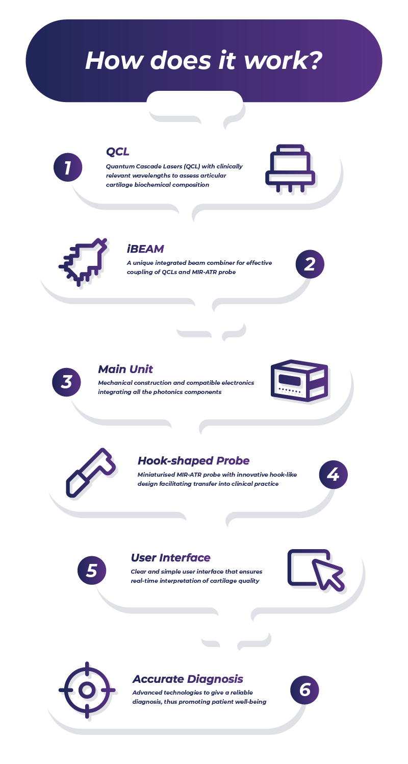
MIRACLE device will measure the biocomposition of cartilage tissue. This can be achieved by using biophotonics, i.e. the combination of biology and photonics. Photonics is an area of study that involves the use of radiant energy (such as light, lasers and so on), whose fundamental element is the photon.
The concept behind MIRACLE is quite simple. Cartilage tissue is made of very specific molecules. Some molecules have particular response when illuminated with a specific light source (as for example specific lasers). This phenomenon is called molecular vibration and it can generate a frequency spectrum, which works as a molecular ‘finger print’ for the cartilage tissue. Healthy and damaged cartilage have different spectra. Also, different damage levels will show different spectra. Based on these scientific evidences, MIRACLE is assembling a device capable of compiling this information into a colour-coded map of healthy and damaged tissue during the arthroscopy procedure. The surgeon will then have all detailed information in a graphical user interface. The device is not capable of doing external examination and will be used only during arthroscopy.