Arthroscopy, The Journal of Arthroscopic and Related Surgery webpage has launched several podcasts discussing the new releases and trends in the scientific field of arthroscopy.
On the 7th of December the episode number 91 was released, discussing the manuscript “Predicting Severe Cartilage Damage In the Hip: A Model Using Patient-specific Data from 2,396 Hip Arthroscopies”, authored by Dr. Marc Philippon. The episode was podcasted by Justin Arner and brings an interesting discussion on the work developed and the new ways to go.
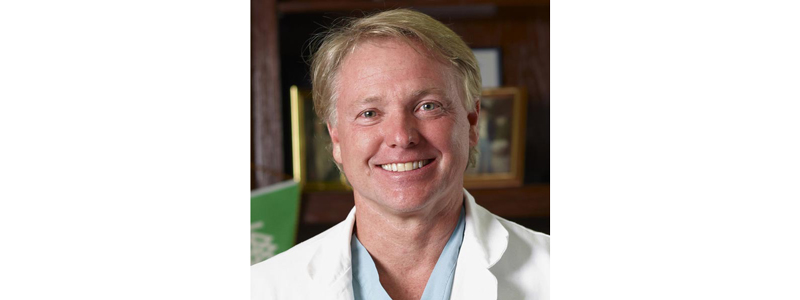
Dr. Marc Philippon is a recognized hip surgeon, internationally known for performing joint preservation techniques utilising arthroscopic hip surgery to treat painful joint injury in high-level athletes who constantly use powerful hip rotation. He has successfully treated close to 1000 Professional and Olympic athletes with many of them returning to high performance.
As a Key Opinion Leader on the matter of arthroscopy and in the treatment of joint injury, through innovative procedure and treatments, Dr. Philippon was invited to discuss his latest published work entitled, “Predicting Severe Cartilage Damage In The Hip: A Model Using Patient-specific Data From 2,396 Hip Arthroscopies,” which was published in the July 2019 issue of the Arthroscopy Journal.
In the podcast Dr. Philippon talks about the development of the aforementioned study that led to the published issue, and how he decided to study and work on an algorithm to predict severe cartilage damage in the hip. The important take out, is the importance of the quality of the technique enclosed to the procedure.
Several questions about the procedure applied by the Doctor in the study were discussed, including the application of preoperative magnetic resonance imaging (MRI), and the combination between MRI and 3D methods. Dr. Philippon highlights that the access to MRI in the different regions in the world are not exactly the same, but for sure, MRI itself is very important “to see in more detail the bone marrow and periarticular musculature, tendonitis and joint effusion”, but still, only MRI is not enough to have an accurate diagnosis or a good prediction on these types of injuries.
When questioned about the application of three-dimensional studies combined with computed tomography (CT) or MRI, Dr. Philippon answered that three-dimension problems required three-dimensional studies and highlights the importance on the studies combining arthroscopy and 3D technologies such as MRI, which by itself is not efficient to detect cartilage damage, to better define the extension of lesion in the joint and to be more precise in the clinical procedure – “…important to take both into consideration and, certainly, the 3D analysis from CT or MRI is certainly a very important adjunct to our preop planning.”
The MIRACLE project is an excellent example of a new and effective arthroscopy MIR-ATR imaging system that enables a real-time and in-depth examination of the articular cartilage, unlike the other technologies in the market that require imaging techniques like CT or MRI to get better prediction on the cartilage injuries. The MIRACLE equipment provides a biochemical composition, which enables the orthopaedic surgeons to have an accurate evaluation of the quality of the cartilage and the extension of injuries or diseases in the joints, mainly in the diagnosis of degenerative joint diseases such as osteoarthritis, during arthroscopy surgery, with no need of applying other technologies.