MIRACLE: “Mid-infrared arthroscopy innovative imaging system for real-time clinical in-depth examination and diagnosis of degenerative joint diseases”
The Miracle Project aims to transfer a well-established technology, the Mid-infrared Spectroscopy, into clinical use. Specifically, the project proposes to use this technology in the development of a Real-Time Diagnostic tool for cartilage lesions.
Mid-infrared spectroscopy is an effective and widely used sample characterization and analytical technique. The application of this technology to the in vitro diagnoses of cancer has already been published and is presented as a highly promising technique, which will surpass the lack of highly sensitive and specific methods for early detection of cancer. This technology is also described as “simple, rapid, accurate, inexpensive, non-destructive and suitable for automation compared to existing screening, diagnosis, management, and monitoring methods” and that will “potentially improve clinical decision-making and patient outcomes by detecting biochemical changes in cancer patients at the molecular level” (1). An in-progress clinical trial is testing the applicability of this technology for the diagnostics of chronic endometritis (2). It started in 2019 and is expected to end in 2022. This shows how updated the application of the technology is to the clinics.
The clinical use of MIR-spectroscopy is a recent and promising field of research. However, clinical translation presents major challenges, such as the downsizing of the equipment.
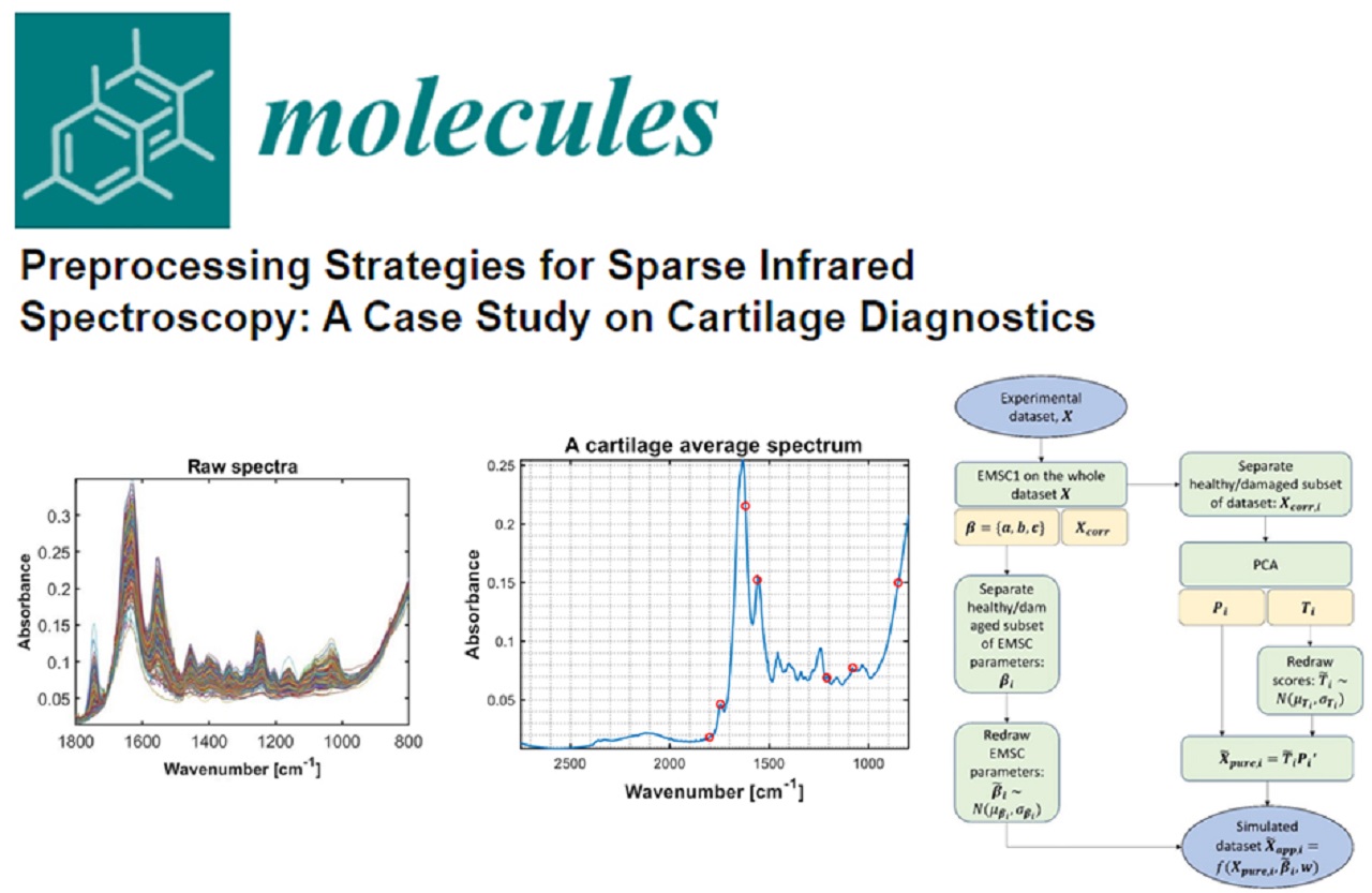
The design of the Miracle Project was based on previously obtained data by the OULU partner, demonstrating the feasibility of the MIR-based method for monitoring compositional changes on articular cartilage. The project partners accepted the challenge to transfer this knowledge from the laboratory bench to clinical use. This implies several technical challenges involving tailored laser light sources, beam combiners, waveguides (for spatial resolution), and integration of each optical component.
The medical device proposed in the MIRACLE Project is intended to be used during arthroscopy (a minimally invasive surgery) and will provide data on the biochemical composition of the articular cartilage. It represents a major advance in the diagnosis of cartilage lesion, which is, nowadays, based on visual inspection and manual probing of the cartilage tissue, that is highly subjective and of poor repeatability.
Additionally, the Project proposes to develop a user-friendly software that will integrate the data from the spectroscopy and convert it into meaningful analytical parameters. The surgeons, even inexperienced in spectroscopy, will be able to understand the data, as it will be converted in a colorimetric scheme that, in real-time, indicates the quality of the cartilage.
The Miracle device will bring to the arthroscopy market the first MIR-based probe providing an accurate and quantitative diagnostic tool for cartilage lesions. Moreover, the designed probe might be applied to evaluate the chemical composition of other soft tissues, such as tendons.
Although this is not a hidden secret, we are convinced that this is a powerful technology that deserves to be explored for application in numerous clinical conditions and will represent a major advance in real-time diagnosis.
(1) Su, K. Y. and W. L. Lee (2020). “Fourier Transform Infrared Spectroscopy as a Cancer Screening and Diagnostic Tool: A Review and Prospects.” Cancers (Basel) 12(1).


