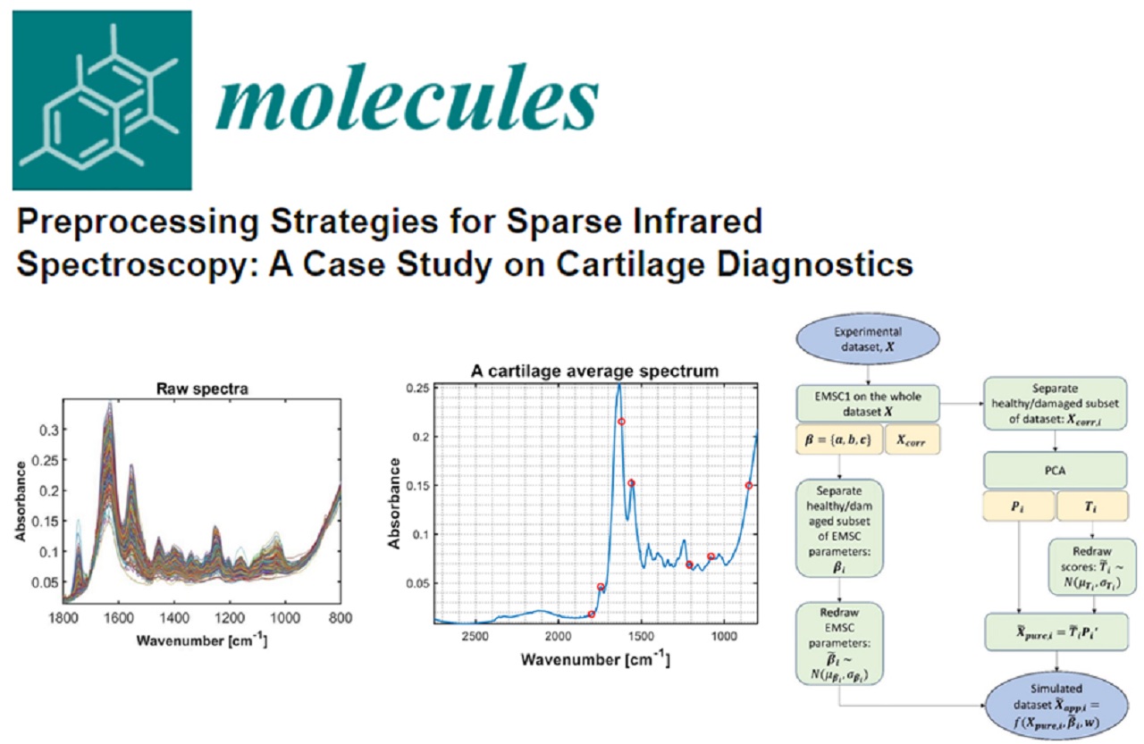
Short summary of the presentation held by Dr. Harold Brommer, Faculty of Veterinary Medicine, Utrecht University, The Netherlands, at the MIRACLE Project Symposium, “A Bright New Future for Arthroscopy”, EORS Congress, in Rome, 15th of September, 2021.
Articular cartilage is a very delicate tissue that, once destroyed, cannot be repaired. Currently, arthroscopy is the gold standard for assessment of the integrity of the surface and the functionality of the tissue.
The current practice of arthroscopy consists of looking at the surface and probing of the tissue with a metallic hook probe. However, no comprehensive assessment of the entire cartilage layer can be achieved, it is only possible to visualize the articular surface of the cartilage layer. Moreover, assessment of functional properties is subjective leading to a poor interobserver agreement making a decision during surgery also subjective.
There are a number of arthroscope-guided intra-articular diagnostics such as spectroscopy, ultrasonography and optical coherence tomography. Ultrasonography and optical coherence tomography both give information of the integrity of the cartilage layer. Spectroscopy reflects the biochemical composition of articular cartilage and, related to that, the functional properties. Cartilage lesions will lead to changes in biochemistry that can be detected by changes in the derivative infra-red spectra.
There are two types of spectroscopy: near infra-red and mid-infrared. Near infra-red spectroscopy is performed at a wavelength of 780-1000 nm and reflects the entire cartilage layer up to subchondral bone. Mid-infrared spectroscopy is performed at a wavelength of 1000-3000 nm and only reflects the upper 20-30 µm of the cartilage layer.
Early diagnosis of articular cartilage degeneration is desirable at a stage that there is softening of the tissue without any macroscopical damage. In that stage initial biochemical changes such as proteoglycan depletion and water uptake has taken place which is reversible as long as there is no damage of the collagen network. This initial stage of cartilage damage can potentially be detected using arthroscopic mid-infrared spectroscopy.
There is currently no functional hook probe available for clinical mid-infrared spectroscopy, but recently we have gained clinical experiences with near-infrared spectroscopy. In an experimental study in which cartilage lesions were made in the stifle joint of Shetland ponies, repair and changes of the surrounding cartilage was evaluated post-operatively using near-infrared spectroscopy. It turned out that biomechanical/functional properties of articular cartilage could adequately be predicted from near-infrared spectra both at locations nearby the created lesions as well as at locations further away. Moreover, the functional properties of cartilage in these joints were different compared to controlled joints. These findings substantiated the clinical value of arthroscopic spectroscopy that functional properties could be objectively measured in vivo.
With the use of mid infra-red spectroscopy, it is foreseen that objective assessment of invisible cartilage damage can be performed in a joint that is prone to the development of osteoarthritis, such as meniscal lesions, cruciate ligament lesions or traumatic osteo-chondral lesions. That mean that very early detection of osteoarthritis is feasible in the near future. This may have consequences not only for the prognosis of joint trauma, but also for post-operative medication and rehabilitation programmes. Altogether, a big step forward is anticipated in the near future regarding a better management of osteoarthritis, saving costs and a better patient well-being and quality of life.


