
Osteoarthritis disease burden is similar to rheumatoid arthritis
Another indication of the urgent need for improved treatments and strategies for prevention of osteoarthritis came out this week.
Laser packaging solution for QCL light source
Nanoplus is developing quantum cascade lasers (QCLs) for the MIRACLE project. These lasers will be used to determine the biochemical composition of joint cartilage.
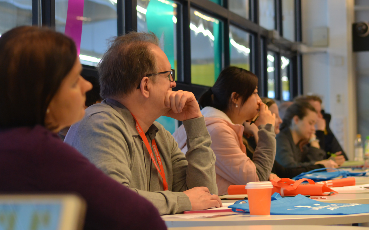
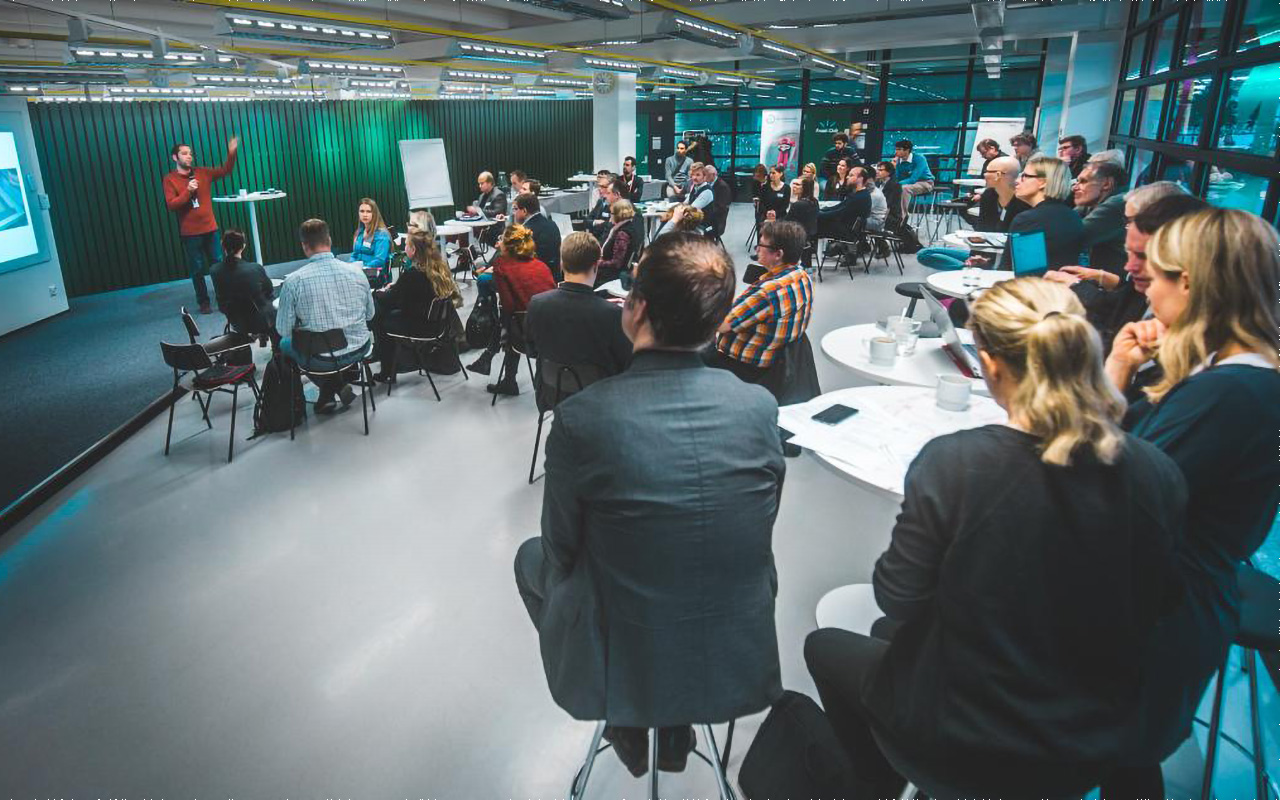
BioPhotonics for Healthcare workshop succeeded
The BioPhotonics for Healthcare Workshop organized by the MIRACLE consortium took place on 13th and 14th of March 2019 at the Tellus Stage, University of Oulu, Finland.
Selection of specific wavelengths for osteoarthritis detection
For the MIRACLE project, laser sources in the mid-infrared range will be used to investigate characteristic peaks in the absorption spectrum of articular cartilage.

CEN webinar “Good practices for the development of harmonised standards under the Medical Device Directive”
On February 26th of 2019 a CEN Webinar about Good practices for the development of harmonised standards under the Medical Device Directive.

MIRACLE celebrates first year of project
The MIRACLE project reached the end of its first year and to mark that occasion, a project meeting was held in Ulm to discuss the latest developments and to plan the activities for the coming months.
MIRACLE organises BioPhotonics for Healthcare workshop
The MIRACLE consortium is organizing the BioPhotonics for Healthcare workshop which is taking place on the 13thand 14thof March at the Tellus Stage, University of Oulu, Finland.

News on standardization for 2019
New year always comes with new purposes and objectives. 2019 has started with some important novelties related to standardization:

Data modelling as medical decision support system for arthroscopy surgery
Artificial Intelligence (AI) and data modelling are becoming part of our everyday life bringing comfort and simplifying our decisions.

Reasons demanding new cartilage diagnosis tools
Currently, damage to the articular cartilage is diagnosed by standard imaging techniques (X-Ray, Computed tomography or MRI) or using more invasive methods such as manual probing during arthroscopy.